
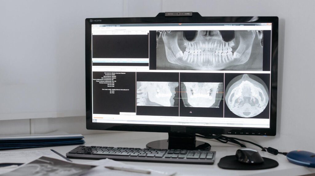
Losing a tooth does more than change your smile. When the tooth and its root are gone, the jawbone that depended on that root starts to weaken and shrink.
The bone around the extraction site loses mechanical stimulation and begins a resorption process. You might notice bone loss within weeks, and the first year can bring significant volume loss.
Let’s talk about how the alveolar bone depends on chewing forces, why that signal disappears after extraction, and what you can do—like implants or grafting—to protect your bone and future options. Understanding how missing teeth lead to jawbone loss is the first step to deciding how to preserve what you have. I’ll walk you through the biology, the timeline of changes, and some practical steps to help you make decisions that actually matter.
Jawbone Anatomy and Function
The jawbone holding your teeth is pretty specialized. It’s built for support, sensation, and handling force.
It keeps adapting to chewing loads and to whether tooth roots are present. That’s a lot of work for a bone.
Structure of the Alveolar Bone
The alveolar bone forms the tooth sockets (alveoli), sitting right on top of the basal jawbone. It’s got a dense cortical plate on the outside and a spongy trabecular core inside.
The cortical plate gives the bone its rigidity and a surface for the periodontal ligament to attach. The trabecular bone absorbs and spreads out chewing forces.
Tiny channels in the alveolar bone carry blood vessels and nerves to nourish the bone and the periodontal ligament. The bone around each root is often just a few millimeters thick, so it reacts quickly to changes in load or inflammation.
Lose a root, and you lose local mechanical stimulation. The bone in that spot starts thinning almost immediately.
Role in Tooth Support
Your alveolar bone’s main job? Anchoring teeth with the periodontal ligament (PDL). Collagen fibers from the PDL stick into both the tooth cementum and the alveolar bone, making a shock-absorbing connection.
This setup lets teeth safely transmit forces into the jaw when you chew. When the PDL senses load, it sends out signals that keep bone density up in that area.
Remove a tooth, and those signals vanish. No PDL, no root—so the bone there doesn’t get its usual maintenance and begins to resorb.
Bone Remodeling Processes
Bone remodeling is always happening, balancing breakdown by osteoclasts and building by osteoblasts. Mechanical loading boosts osteoblast activity and bone growth.
Less load or more inflammation means more osteoclast action and bone loss. Remodeling gets regulated by signals like RANK/RANKL/OPG, prostaglandins, and local growth factors.
These signals respond to chewing, tooth movement, and healing after extraction. After tooth loss, the balance tips toward resorption at the old socket, leading to a visible drop in bone width and height over time.
Mechanisms of Bone Loss After Extraction
Jawbone shrinks because the tooth root’s mechanical stimulation disappears, and the biology shifts toward resorption. Timing, cellular signals, and your own risk factors all play a part in how much bone you lose and how fast.
Biological Drivers of Resorption
When you lose a tooth, the alveolar bone stops getting mechanical load through the root. Osteoblasts (the bone builders) do less, while osteoclasts (the bone breakers) get busier.
This imbalance leads to bone loss around the extraction site. Inflammation from surgery or leftover infection bumps up cytokines like IL‑1 and TNF‑α, which encourage osteoclasts even more.
Blood clots and soft-tissue healing change the local blood supply and growth-factor signals, which also affect bone remodeling. If you don’t get a load-bearing restoration (like a bridge, denture, or implant) soon enough, the bone leans toward resorption instead of rebuilding.
Socket preservation or grafting can help by giving the bone a scaffold and signaling molecules to keep osteoblasts working.
Timeline of Jawbone Changes
Bone remodeling kicks in just days after extraction. In the first week, you’ll see clot organization and early osteoclast activity at the socket walls.
You probably won’t notice much change at first, but resorption starts right away. Between 4–8 weeks, the socket fills with woven bone, but the ridge width starts shrinking.
Most of the horizontal loss happens in the first three months. By 6–12 months, you could lose up to about 25% of ridge width, with slower vertical shrinkage continuing after that.
If you’re considering an implant, early action—within weeks or a few months—matters most for keeping the bone’s width and shape. Wait too long, and you’re more likely to need ridge augmentation to rebuild lost volume.
Risk Factors Influencing Bone Loss
Local factors like large extraction sockets, missing neighboring teeth, gum disease, and infection speed up resorption. Sockets with thin buccal bone are especially at risk for quick horizontal collapse.
Systemic stuff matters too: smoking, uncontrolled diabetes, osteoporosis, and some meds (like long‑term steroids) slow healing and boost osteoclastic activity. Age plays a role—older patients usually form new bone more slowly.
Treatment choices count. Traumatic extractions, skipping socket preservation, or waiting too long for a prosthetic raise your risk of losing a lot of ridge. Gentle extraction, grafting, and timely implants can help keep your bone.
Preventing and Managing Post-Extraction Bone Loss
Acting early helps preserve the ridge shape, keeps implant options open, and reduces long-term facial changes. What you do at the time of extraction really determines how much bone you’ll keep.
Socket Preservation Techniques
Socket preservation means putting a barrier or filler in the empty socket right after extraction to slow bone loss. Usually, your dentist will pack in a bone substitute (allograft, xenograft, or synthetic), cover it with a resorbable collagen membrane, and maybe add a few sutures.
This approach cuts down on horizontal and vertical bone loss over the first year and makes future implants easier. The whole thing usually adds 10–30 minutes to the extraction and heals in about 4–6 months before you move on to final restoration.
Some quick points:
- Materials: you’ll see allograft (human), xenograft (bovine), or synthetic (beta-TCP, HA).
- Membranes: resorbable collagen is typical, but sometimes a nonresorbable one is used for extra stability.
- Healing: most grafts integrate in 3–6 months, depending on the site and material.
Dental Implant Solutions
Immediate or early implant placement puts a “root” back in the bone and restores mechanical stimulation, which helps stop further resorption. You can get an implant the same day as extraction, within weeks, or after the socket heals.
The timing depends on things like infection, bone volume, and soft-tissue health. Immediate implants do best when the socket walls are intact and there’s no infection.
If there are gaps between the implant and socket walls, your dentist will add bone graft material or do ridge augmentation. For implants to work, you need good oral hygiene, enough bone, and well-managed health conditions—so if you smoke or have diabetes, it’s worth getting those under control.
Bone Grafting Procedures
When you’ve lost bone in the socket or along the ridge, bone grafting can rebuild the area before or during implant placement. The main options—particulate grafts, block grafts, and ridge-splitting—depend on the defect’s size and where it’s located.
Particulate grafts work well for small deficiencies. They usually integrate within a few months.
Block grafts, which use your own bone (often from the hip or chin), add bulk for bigger horizontal or vertical defects. Of course, these need a second surgical site, which not everyone loves.
Ridge-splitting and guided bone regeneration involve membranes and screws to shape the new ridge. It’s a bit technical, but sometimes that’s the only way.
Healing takes a while—anywhere from four to nine months for the bone to mature enough. That’s not a quick fix, but it’s usually worth it.
Risks? Sure, there are some: graft failure, infection, or the bone not sticking around as much as you’d hoped. Careful planning and a sterile setup help a lot, but nothing’s ever guaranteed.
Costs and complexity go up as the reconstruction gets bigger. If grafting isn’t a good fit, maybe look at alternatives like hybrid prostheses. It’s always a conversation worth having.